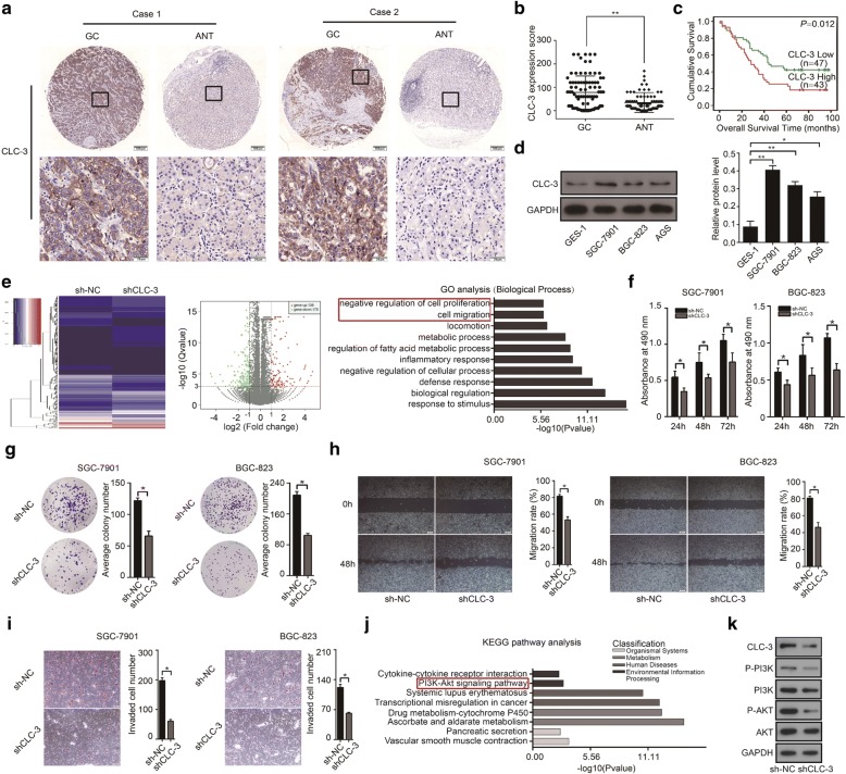
Fig. 1.
Overexpression of CLC-3 was a poor prognostic biomarker for GC patients, and CLC-3 knockdown inhibited cell proliferation and migration in vitro. a Representative images of CLC-3 expression in GC tissues and adjacent normal tissues (ANT). b The expression of CLC-3 in 90 GC tissues was higher than that in ANT. c High expression of CLC-3 was associated with poor prognosis in GC patients. d The basic protein expression of CLC-3 in human normal gastric epithelial cells (GES-1) and human GC cell lines (SGC-7901, BGC-823, and AGS). e The heatmap and volcano plot of RNA sequencing were constructed after the CLC-3 knockdown. Cell proliferation and migration were identified as the primary biological functions of CLC-3 according to the Gene Ontology (GO) analysis. f, g Knockdown of CLC-3 inhibited the proliferation and clonogenicity of SGC-7901 and BGC-823 cells (n = 3). h, i Knockdown of CLC-3 inhibited the migration and invasion of SGC-7901 and BGC-823 cells (n = 3). j The PI3K/Akt signaling pathway was enriched according to the Kyoto Encyclopedia of Genes and Genomes (KEGG) pathway analysis of CLC-3 knockdown. k Knockdown of CLC-3 reduced the levels of key targets in the PI3K/Akt signaling pathway. *P < 0.05, **P < 0.01

