Fig. 7.
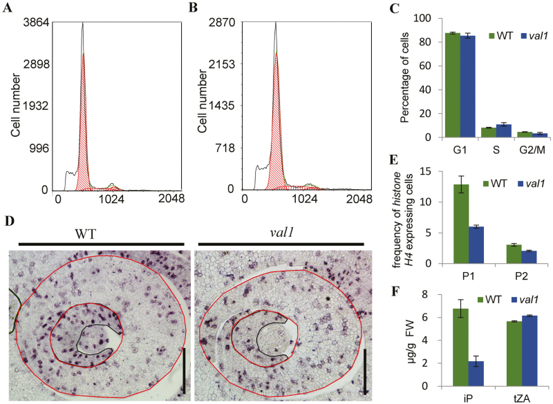
Involvement of VAL1 in cell division in leaves. (A, B) Flow cytometry measurements of nuclei from the shoot apex in 10-d-old wild-type (WT) (A) and val1 plants (B). (C) Quantification of the DNA profiles of WT and val1 plants. (D) Expression of histone H4 in cross-sections of the shoot apical meristems in the WT and val1 at the tiller stage. Black and red lines represent P1 and P2 primordia, respectively. Scale bars are 100 µm. (E) Frequency of histone H4-expressing cells in the leaf primordia indicated by red circles in (D). P1, P1 promordium; P2, P2 promordium. (F) Contents of isopentenyladenine and trans-zeatin in the WT and val1 mutant seedlings. Data are means (±SD) of three biological replicates.

