Fig. 3.
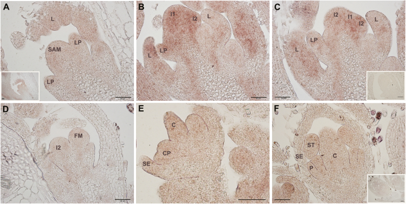
In situ localization of MtSOC1a transcripts through the floral transition in longitudinal sections of wild-type R108 plants grown in LDs, after 1 week of vernalization. (A) Vegetative shoot apical meristem (SAM) with leaf primordium (LP) and leaf (L). The inset shows the same section at a lower magnification. (B and C) SAM at the initial reproductive stage. Increased transcript abundance was detected in SAM, L, LP, and primary (I1) and secondary (I2) inflorescence meristems. (D–F) Weak MtSOC1a expression was detected during early stages of floral meristem development (D), during differentiation of floral organs (E), and in young flower buds (F). No expression was detected using a sense control MtSOC1a probe (inset images in C and F). C, carpel; CP, common primordium for petal and stamen; P, petal; SE, sepal; ST, stamen. The black scale bar represents 50 µm.

