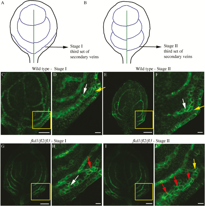
Fig. 9.
PIN1–GFP expression in the developing third set of secondary veins in the wild type and fkd1/fl2/fl3 triple mutant. (A and B) Diagram of successive stages of development of the third set of secondary veins; lines indicate PIN–GFP expression domains (PEDs). (C–F) PIN1–GFP expression in the third set of secondary veins of the wild-type stage I (C, D) and stage II (E, F) first leaf (boxed area in C and E enlarged in D and F, respectively). (G–J) PIN1–GFP expression in the third set of secondary veins of the fkd1/fl2/fl3 triple mutant stage I (G, H) and stage II (I, J) first leaf (boxed area in G and I enlarged in H and J, respectively). White arrows indicate basal PIN1–GFP, yellow arrows indicate lateral PIN1–GFP, and red arrows indicate PIN1 localization on all sides (scale bar=10 μm).

