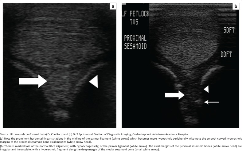
FIGURE 4.
(a) Transverse ultrasound scans performed at the level of the palmar proximal sesamoid bones of the left forelimbs of a normal palmar ligament and sesamoid bones of an adult mixed breed horse (b) and a limb affected by axial sesamoiditis and palmar desmitis in an adult Thoroughbred horse. Lateral is to the left of the images.

