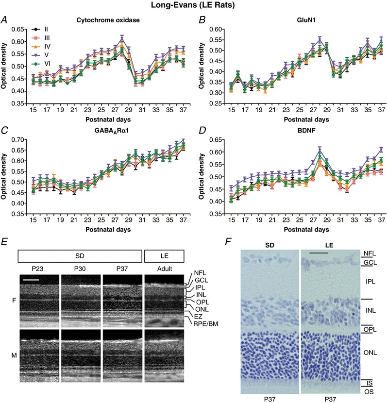
Figure 8. Developmental comparisons between Long Evans and Sprague‐Dawley rats at the cortical and retinal levels.

A–D, neurochemical development of layers II to VI visual cortical neurons in LE rats. A 1‐day delay in eye opening in these rats led to analysis from P15 to P37. The developmental trends of CO (A), GluN1 (B), GABAARα1 (C) and BDNF (D) in LE rats were virtually identical to those of SD rats (Fig. 6). N = 50–100 neurons from two litters for each layer at each time point tested. E, OCT of SD and adult LE rats. No evidence of major retinal changes was observed in SD rats from the third to fifth postnatal weeks. Log intensity OCT images from the same female (F) and male (M) SD rats at P23, P30 and P37 are shown. For comparison, images from an adult female and an adult male LE rats are included at approximately the same retinal location. For display purposes, images have been manually contrast stretched. Scale bar: 100 μm. F, Nissl‐stained retinal sections from SD and LE rats, both at P37. All retinal layers were comparable in thickness between the two strains. NFL, nerve fiber layer; GCL, ganglion cell layer; IPL, inner plexiform layer; INL, inner nuclear layer; OPL, outer plexiform layer; ONL, outer nuclear layer; EZ, ellipsoid zone of the inner segment of photoreceptor cells; IS, inner segment; OS, outer segment of photoreceptors; RPE/BM, retinal pigmented epithelium/Bruch's membrane. Scale bar: 25 μm.
