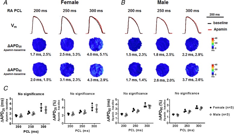
Figure 1. Effects of I KAS blockade on APD at basal condition.

A and B, representative V m traces, ΔAPD25 and ΔAPD80 maps in female (A) and male (B) rabbit ventricles at RA PCL 200, 250 and 300 ms under Protocol I. The apamin‐induced APD prolongation was less than 5% in the absence of β‐adrenergic stimulation. Representative V m traces were obtained at the LV base. C, summary data showed that no significant difference existed in ΔAPD25 and ΔAPD80 between females and males at baseline (by two‐way ANOVA with Sidak's post hoc test). [Color figure can be viewed at http://wileyonlinelibrary.com]
