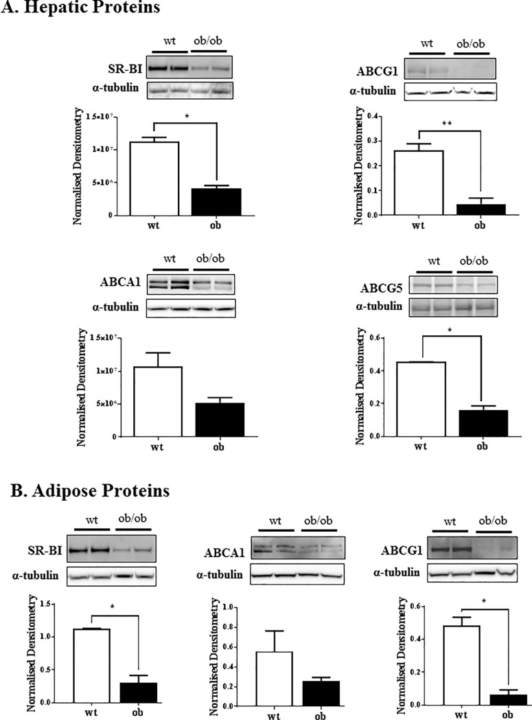
Fig 3. Western blot for SR-BI, ABCA1, ABCG1 and ABCG5 protein expression.
Liver (A) and adipose (B) tissue were homogenised into lysates in RIPA buffer with protease inhibitors and PMSF. The lysates (10–20 μg protein for liver and 40 μg protein for adipose) were loaded onto a 3–12% SDS gel and transferred onto nitrocellulose membrane. The membranes were probed for SR-BI, ABCA1, ABCG1 and ABCG5 (liver only) and visualised with ECL and densitometry. Results are normalised to the α-tubulin loading control. n = 3–4 mice of each genotype; *p<0.01.

