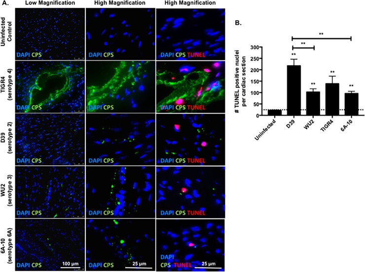
Fig 3. TUNEL staining reveals cardiomyocyte death close to TIGR4, but dispersed damage in hearts from mice challenged with other strains.
A) Representative low and high magnification immunofluorescent microscopy images of cardiac sections form mice infected with TIGR4, D39, WU2, and 6A-10 pneumococci 30 hours post infection. The heart sections were probed for S. pneumoniae (green), nuclei (blue, DAPI) and fragmented nuclei (red, TUNEL probe). B) Enumeration of TUNEL positive nuclei per cardiac section of mice infected with TIGR4, D39, WU2, and 6A-10 pneumococci 30 hours post-infection. For each strain, 4–6 sections were examined. Statistical analyses were performed using a non-parametric one-way ANOVA. P value: ** ≤ 0.01, **** < 0.0001; data is represented as mean ± SEM.

