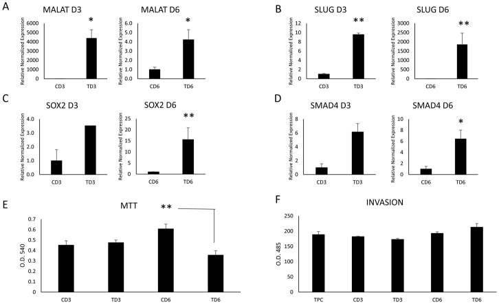
Fig. 3.
RT-qPCR results of naïve TPC-1 cells incubated with 1 × 10⁸ control and TGFβ treated TPC1 exosomes isolated at day 3 and day 6 of treatment. (A) LncRNA MALAT1 was significantly upregulated in the naïve TPC-1 cells by TGFβ treated exosomes on both days analyzed. The EMT factor SLUG (B), stem cell marker SOX2 (C) and the TGFβ pathway protein SMAD4 (D) were upregulated only on day 6 by TGFβ treated exosomes. (E) Proliferation of naïve TPC-1 cells incubated with control and TGFβ treated TPC1 exosomes was measured by MTT assay. Day 6 TGFβ treated exosomes significantly reduce proliferation. (F) Invasive properties of naïve TPC-1 cells incubated with days 3 and 6 control and TGFβ treated TPC1 exosomes were analyzed. No significant difference was seen, however, day 6 TGFβ treated exosomes began to increase invasive potential of the naïve TPC-1 cells. Data represented by means +/− SEM. * = p<0.05, ** = p<0.01.

