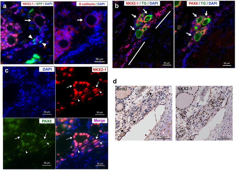
Figure 2. Formation of thyroid follicles from NKX2-1-positive non-follicular cells.
(a) Immunofluorescence for NKX2-1 and GFP (left), and E-cadherin (right) in the Nkx2-1(fl/fl);TPO-cre mice that received bone marrow cells from GFP-transgenic mice. Arrows: a newly formed follicle connected to a zone of cells containing many NKX2-1-positive cells. Arrowheads: GFP-positive cells. (b) Immunofluorescence for NKX2-1 and TG (left), and PAX8 and TG (right). Arrows: newly formed follicles located in the middle of a zone of cells containing many NKX2-1 positive cells, but very weak PAX8 expression (shown by bars). (c) Immunofluorescence for NKX2-1 and PAX8 near a newly formed follicle. Arrows: NKX2-1 and PAX8 both strongly expressed. Arrowheads: PAX8 expression is weaker than NKX2-1, resulting in stronger magenta color in the merged image. (d) Immunohistochemistry for BrdU (left) and NKX2-1 (Right). Arrows: BrdU positive cells.

