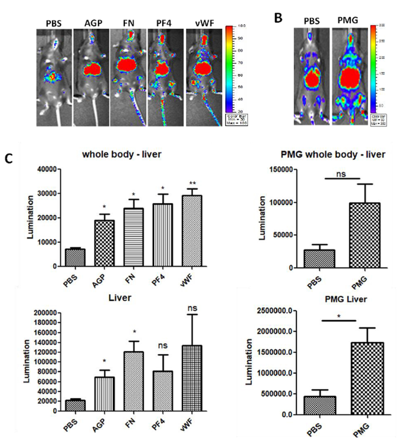
Fig. 4. Other serum protein enhanced the global transduction of AAV9 vectors.

1×10¹⁰ vg of AAV9/luc were incubated with either AGP, FN, PF4, vWF, or PMG at the normal physiological concentration for 2 hours at 4 °C and then injected into C57BL/6 mice via the retro-orbital vein. Imaging was taken at day 7, and the photon signal was measured and calculated. The data represents the average and standard deviation from 5 mice. The asterisk indicates the significant difference (p<0.05).
