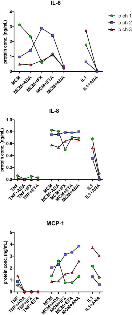
Figure 4. Cytokine secretion by chondral microspheroids after their exposure to inflammatory factors and anti-inflammatory biological drugs.
Quantities of IL-6, IL-8 and MCP-1 (ng/mL) detected in supernatants of microspheroid chondral tissues formed from human OACs (p ch1, p ch2 and p ch3) following their 24 h exposure to TNF-α, MCMh.s. working solution or IL-1β alone or combined with individual anti-inflammatory biological drugs, adalimumab (ADA), infliximab (IFX), etanercept (ETA) or anakinra (ANA). Single measurements were carried out in pooled supernants of each differently treated microspheroid group. Please, note differences in scales.

