Fig. 2.
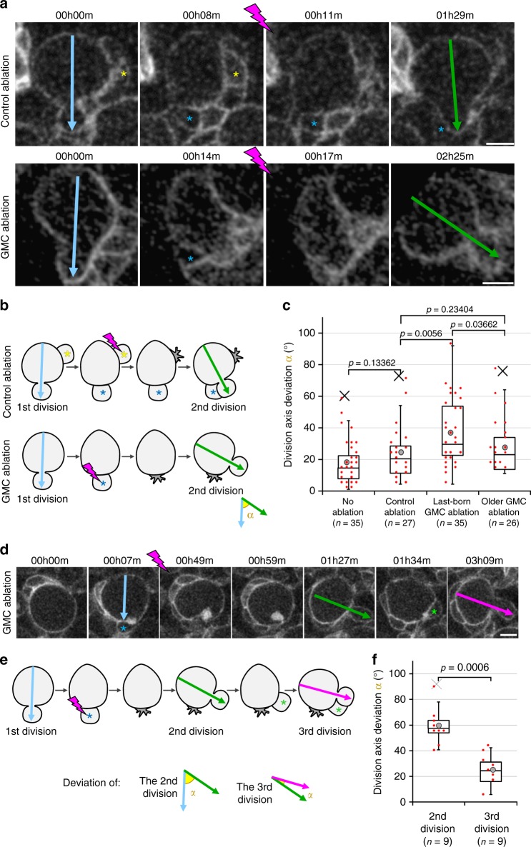
GMC ablation disrupts division axis maintenance. a Two successive divisions of NBs expressing worniu-GAL4-driven PH::GFP, the second division following the control ablation (upper panels) of a NB-neighbouring cell (yellow asterisk) away from the last-born GMC (blue asterisk) or following GMC ablation (lower panels) are shown. The ablation takes place between the two frames separated by a lightning symbol. Arrows: division axis. b Schematic of the ablations and angle measurements. c Deviation of the division axis following no ablation (Average angle: 18 ± 14°, n = 35), control ablation (23 ± 16°, n = 27), last-born GMC ablation (36 ± 20°, n = 35; also shown in Figs. 3c and 5c) or older GMC ablation (27 ± 16°, n = 26). Data from ten independent experiments. For examples of control or last-born GMC ablations resulting in a high misalignment of the second division axis, see Supplementary Movies 6 and 7. d Three successive divisions of NBs expressing worniu-GAL4-driven PH::GFP. The ablation takes place between the two frames separated by a lightning symbol. Blue asterisk: GMC prior to ablation, green asterisk: new last-born GMC. Arrows: division axis. e Schematic of the angles α measured from the movies shown in the previous panel. f Deviation α of the second (59 ± 14°, n = 9) and third (25 ± 11, n = 9) divisions described in the two previous panels. Data from five independent experiments. Scale bar in all panels: 5 µm

