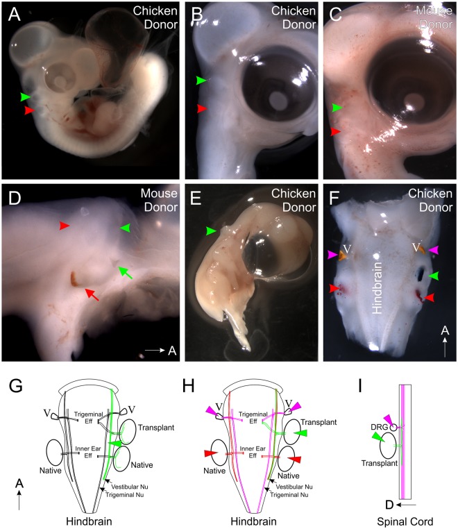
Figure 1.
Transplantations and dye labeling. (A) Chicken embryo with a donor chicken ear (green arrowhead) rostral to the native ear (red arrowhead) following two days of incubation. (B) Chicken embryo with a donor chicken ear (green arrowhead) rostral to the native ear (red arrowhead) following four days of incubation. (C) Chicken embryo with a donor mouse ear (green arrowhead) rostral to the native ear (red arrowhead) following five days of incubation. (D) Side view of dissected chicken head revealing a second external opening (green arrow) adjacent to the transplanted mouse ear (green arrowhead). The native ear and external opening are marked in red. (E) Chicken embryo with a donor chicken ear (green arrowhead) transplanted caudally to the trunk adjacent to the spinal cord. (F) Dorsal view of dissected chicken head showing placement of lipophilic dye into the transplanted ear (green arrowhead), native ears (red arrowheads), and into the trigeminal ganglia (V, magenta arrowheads). (G) Diagram depicting placement of lipophilic dye into the dorsal alar plate (green wedge) and subsequent neurons that would be labeled (Note: in some instances, the trigeminal ganglion was also labeled given the close proximity of the trigeminal nucleus to the vestibular nucleus (See Fig. 2C)). (H) Diagram depicting placement of lipophilic dye into the native ears (red wedge), transplanted ear (green wedge) and trigeminal ganglia (magenta wedge) and subsequent neurons that would be labeled. (I) Diagram depicting the placement of lipophilic dye into the transplanted ear (green wedge) and into dorsal root ganglia (DRG, magenta wedge) and the subsequent neurons that would be labeled.

