Figure 2.
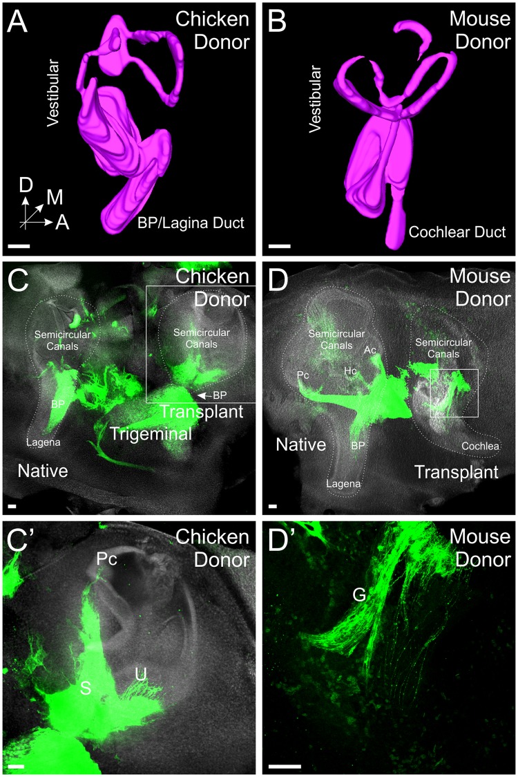
Transplanted ears develop beyond otocyst stage. (A) Three-dimensional (3D) reconstruction of Hoechst staining of a chicken ear three days after transplant. (B) 3D reconstruction of Hoechst staining of a mouse ear four days after transplant. (C) Lipophilic dye labeling (green) from the alar plate and Hoechst (white) staining of a transplanted chicken ear four days after transplant showing innervation of the ears. The basilar papilla (BP, arrow)/lagena grew perpendicular to the plane of the image and is therefore not visible in this image or in E. (D) Lipophilic dye labeling (green) from the alar plate and Hoechst (white) staining of a transplanted mouse ear four days after transplant showing innervation of the ears. Semicircular canals (Ac, anterior, Pc, posterior, Hc, horizontal) are labeled in the native chicken ear. (C’) Higher magnification of C (boxed area) showing innervation of the posterior canal (Pc) utricle (U) and saccule (S) from the transplanted ear. (D’) Higher magnification of D (boxed area) showing otic ganglia (G) adjacent to the mouse ear. Orientation for all panels is as in A (A, anterior, D, dorsal, M, medial). Scale bars represent 100 μm.

