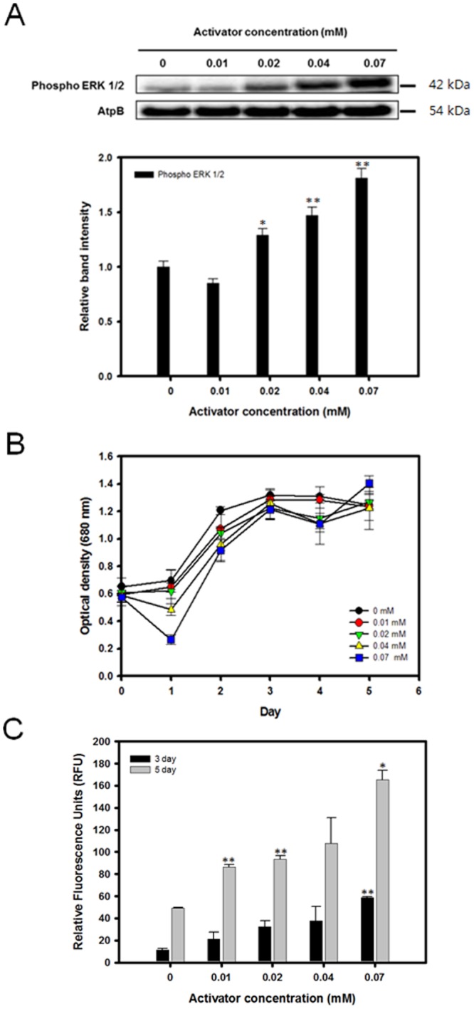
Figure 4.

Expression level of ERK, cell growth profile, and lipid accumulation of C. reinhardtii grown in media containing 0.1 M NaCl with various concentrations of the ERK activator C6 ceramide. (A) Western blot analysis against anti-ERK1/2. ATPβ antibody was detected as a loading control. Full-length blots are presented in Supplementary Fig. S6. (B) Cell growth (O.D. 680 nm). (C) Nile red fluorescence measurement at 3 and 5 days after osmotic stress induction. Data are represented as mean ± SD (n = 3). Significant differences were determined by Student’s t-test and indicated by asterisks (*P < 0.05, **P < 0.01, ***P < 0.001).
