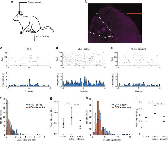
Fig. 5.
Ketamine reduces hyperactivity of neurons in the ACC. a Experimental paradigm for electrophysiological recordings in free-moving rats. b Location of recording electrodes in the ACC. c Raster plots and peri-stimulus time histograms (PSTHs) of representative ACC neurons. Time 0 indicates the onset of noxious (PP) stimulation. d, e Raster plots and PSTHs for CFA-treated rats 5 days after receiving d saline or e ketamine infusion. f, g Chronic pain increased basal firing rates of ACC neurons, but ketamine treatment inhibited this increase. f Histogram showing the distribution of basal firing rates of neurons after saline or ketamine treatment in CFA-treated rats. g Median ± interquartile range for basal firing rates in naive rats, and in CFA-treated rats after ketamine or saline injection; n = 201 (−CFA), 195 (+CFA and saline), and 200 (+CFA and ketamine); p < 0.0001, Kruskal–Wallis test with post-hoc Dunn’s multiple comparisons. See Methods. h, i Ketamine inhibited the enhancement of pain-evoked firing rates of ACC neurons in CFA-treated rats; p < 0.0001. h Histogram showing the distribution of peak firing rates for neurons after saline or ketamine treatment in CFA-treated rats. i Median ± interquartile range for pain-evoked firing rates in naive rats, and in CFA-treated rats after ketamine or saline injection; n = 201 (−CFA), 195 (+CFA and saline), and 200 (+CFA and ketamine); p < 0.0001. Error bars represent S.E.M. Scale bar equals 1000 μm in b. ∗∗∗∗p < 0.0001

