Fig. 1.
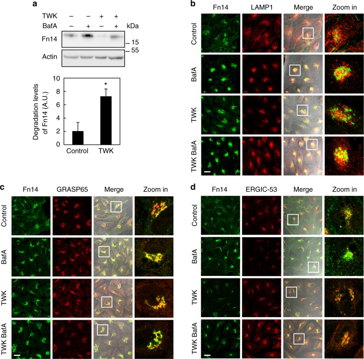
Fn14 is degraded in lysosomes and undergoes trafficking through Golgi. a HeLa cells were treated with BafA and TWK as indicated, extracted proteins were immunoblotted (upper panel) and lysosomal flux was calculated (lower panel) (*P < 0.05, n = 7 biological repeats). b–d Cells were treated as in a, immunostained for Fn14 and LAMP1 (b), GRASP65 (c), or ERGIC-53 (d), and analyzed by confocal microscopy (scale bar, 20 µm). Large magnification of stained cells is presented in the right column. Comparisons by T test; mean ± s.e.m.

