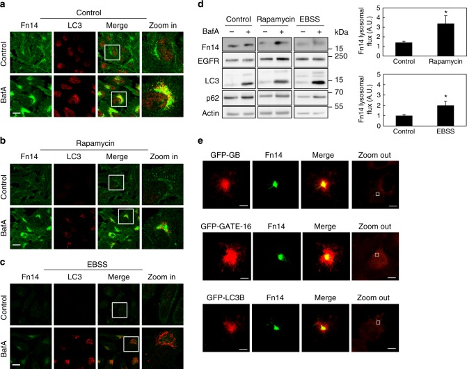
Fig. 3.
Induction of autophagy augments the degradation of Fn14. a–c HeLa cells were incubated for 12 h with BafA as indicated without further treatment (a) or with rapamycin (b) or starved in EBSS (c), immunostained for Fn14 and LC3, and analyzed by confocal microscopy (scale bar, 20 µm). Large magnification of stained cells is presented in the right column. d Proteins extracted from cells treated as in a–c were immunoblotted (left panel) and lysosomal flux was calculated (right panel) (*P < 0.05, n = 2 biological repeats performed in triplicates, comparisons by T test; mean ± s.e.m). e HeLa cells stably expressing GFP-GABARAP (GFP-GB), GFP-GATE-16, and GFP-LC3B were immunostained for GFP and Fn14 and analyzed by 3D STORM (scale bar, 300 nm). Landscape images are presented in the right column (scale bar 10 μm)

