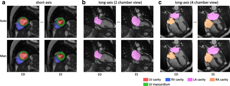
Fig. 3.
Illustration of the segmentation results for short-axis and long-axis images. The top row shows the automated segmentation, whereas the bottom row shows the manual segmentation. The automated method segments all the time frames. However, only end-diastolic (ED) and end-systolic (ES) frames are shown, as manual analysis only annotates ED and ES frames. The cardiac chambers are represented by different colours. a short-axis. b long-axis (2 chamber view). c long-axis (4 chamber view)

