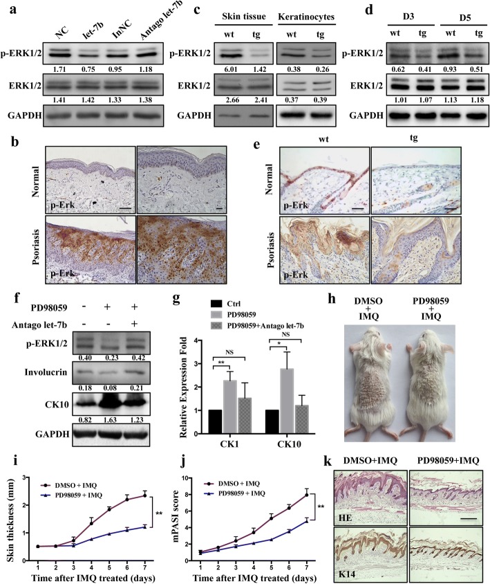
Fig. 6.
let-7b regulated the cell differentiation by reduces p-ERK1/2 signaling in vivo and in vitro. a HaCaT cells were transfected with let-7b mimics or with let-7b inhibitor (“antago let-7b”). Western blotting analysis of phosphorylated ERK1/2 (p-ERK1/2) and total-ERK (ERK1/2). b Immunohistochemistry analysis of p-ERK1/2 analyzed in healthy (n = 4) and psoriasis lesioned skin samples (n = 4). c Western blot analysis shows that p-ERK1/2 is decreased in skin tissue and in primary keratinocytes from keratinocyte-specific let-7b over-expression mice. Expression of total ERK shows no significant difference. Each protein sample results from pools of three mice with the same genotype. d Western blot analysis was performed to detect expression of p-ERK1/2 and total ERK1/2 in transgenic and control mice at days 3 and 5 after treatment with IMQ. e Immunofluorescent staining with p-ERK1/2 (brown) antibodies was performed in normal skin and after treatment with IMQ for 3 days in transgenic and control mice. Scale bars, 100 μm. f, g HaCaT cells were treated with MEK-ERK inhibitors (PD98059: 10 μM) and let-7b inhibitor. The differentiation markers was detected by Western blot (f) and RT-PCR (g). The results obtained analysis were done using t-test. *P < 0.05; **P < 0.01. (h) Phenotypic presentation of mouse back skin for wild-type treated with IMQ and ERK inhibitor (PD98059) for 7 days. The group treated with DMSO is control group. i Skin thickness was measured on the days indicated in the epidermis of mice treated with IMQ or vehicle as well as added PD98059 or DMSO. Symbols represent mean skin thickness ± s.e.m. for five to six mice per group. j mPASI reflecting the intensity of skin inflammation. Two-tailed unpaired Student’s t-test was performed for statistical analysis. Data are shown as means ± s.e.m. k Light microscopy examination of skin sections stained with H&E, or with keratin K14 antibody. Scale bar: 400 μm

