Fig. 7.
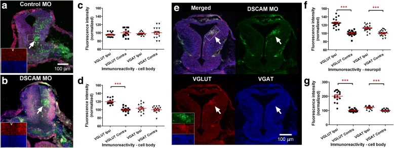
DSCAM downregulation alters excitatory to inhibitory synaptic ratios. a, b Fluorescein-tagged Control MO or DSCAM MO (green) were injected into the light-shaded blastomeres of 4-cell stage embryos; animals were raised to Stage 45. Stage 45 morphant tectal tissues were immunostained with antibodies targeting vesicular glutamate transporter (VGLUT, red) and vesicular GABA transporter (VGAT, blue). Levels of VGLUT and VGAT immunoreactivity were quantified in midbrain regions with MO (right hemisphere-ipsilateral side; white arrows in (a and b) and were compared to the contralateral side (left hemisphere) where MO was not present. Fluorescence intensity for VGLUT (red, top) and VGAT (blue, bottom) immunoreactivities in both hemispheres is also illustrated by the magnified inserts where the ventricle (v) demarcates the separation between the ipsilateral and contralateral sides. c No significant differences in VGLUT or VGAT fluorescence intensity were detected between the ipsilateral side with control MO and the contralateral side without MO. d A significant increase in VGLUT intensity was observed along the cell body layer on the ipsilateral side of the tectum treated with DSCAM MO compared to the contralateral side without MO. f VGLUT and VGAT immunoreactivity was also increased in the neuropil ipsilateral to the DSCAM MO label. e Targeted bulk electroporation was used to focally transfect fluorescein-tagged Control MO or DSCAM MO into the tectum of stage 42 tadpoles; animals were then raised to stage 45 to compare levels of VGLUT and VGAT via immunohistochemistry. The difference in fluorescence intensity in VGLUT (red) and VGAT (blue) immunoreactivity in neighboring areas with and without the DSCAM MO fluorescein tag (green) is illustrated in the overlap and by separating the individual channels (see also the magnified insert; bottom left). g Note brain regions electroporated with DSCAM MO exhibited an increase in VGLUT and VGAT intensity relative to the contralateral non-MO side (Student’s t-test). Error bars indicate mean ± SEM. *** p ≤ 0.001. Scale bars: 100 μm in (a, b, e)

