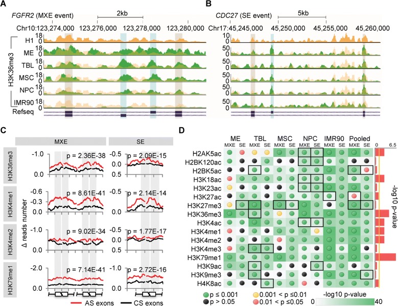
Fig. 2.
Dynamic changes of HMs predominantly occur in AS exons. a, b Genome browser views of representative H3K36me3 changes in MXE (exemplified as FGFR2) and SE (exemplified as CDC27) events, respectively, showing that the changes of H3K36me3 around the AS exons (blue shading) are more significant than around the flanking constitutive exons (gray shading) in 4 H1-derived cell types and IMR90. The tracks of H1 are duplicated as yellow shadings overlapping with other tracks of the derived cells (green) for a better comparison. c Representative profiles of HM changes (normalized Δ reads number) around the AS exons and randomly selected constitutive splicing (CS) exons upon hESC differentiation, shown as the average of all cell lineages pooled together. The ± 150-bp regions (exons and flanking introns) of the splice sites were considered and 15 bp-binned to produce the curves. It shows that the changes of HMs are more significant around AS exons than around constitutive exons, especially in exonic regions (gray shading). The p values, Mann–Whitney–Wilcoxon test. d The statistic significances for changes of all 16 HMs in all cell lineages and pooling them together (pooled), represented as the -log10 p values based on Mann–Whitney–Wilcoxon test. The detailed profiles are provided in Additional file 1: Figure S4. Black boxes indicate the cases that HMs around constitutive exons change more significantly than around AS exons, corresponding to the red-shaded panels in Additional file 1: Figure S4. Sidebars represent the significances whether the changes of HMs are consistently enriched in AS exons across cell lineages, showing the link strength between AS and HMs and represented as the -log10 p value based on Fisher’s exact test. The yellow vertical line indicates the significance cutoff of 0.05. Also see Additional file 1: Figure S4

