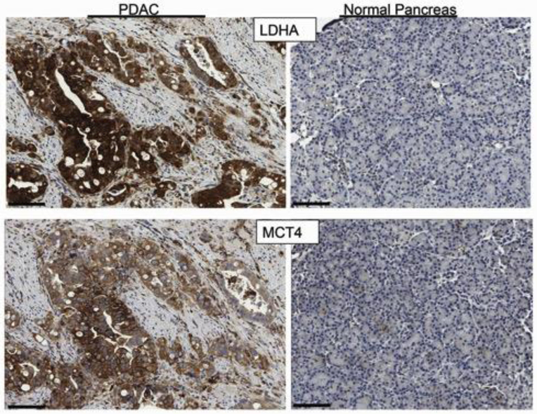
Figure 1.
IHC of IPMN-associated pancreatic adenocarcinoma and a region of normal pancreas. Upper panel) Strong cytoplasmic staining of LDHA was observed in >75% of tumor cells, while normal acinar cells were unstained. Normal ducts exhibited patchy weak staining. Lower panel) Moderate to strong plasma membrane staining of MCT4 was observed in >75% of tumor cells. A few scattered acinar cells and normal ductal cells were weakly or moderately stained. In this particular example, stromal cells adjacent to the adenocarcinoma were predominantly unstained. The normal pancreas core was obtained from a pylorus preserving Whipple procedure. The 70-year-old female patient presented with a mixed MD/BD IPMN and main duct ectasia with associated abdominal pain. There was no evidence of invasion on surgical pathology and the patient is alive 13 years post-resection. Scale bar = 100 microns.

