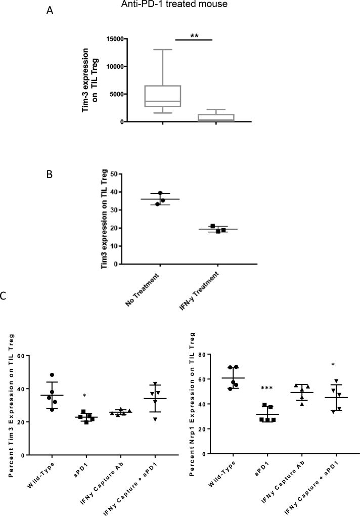
Figure 5. Tim-3 expression is downregulated after PD-1 blockade ex vivo in mouse TIL.
(A) Mice bearing HNSCC tumors were treated with anti-PD-1 or isotype Ab (3mg/kg) for five doses on alternating days from days 12–20. TILs were extracted from anti-PD-1 or isotype mAb treated murine tumors, then Tim-3 expression was tested on TIL Treg by flow cytometry (n=7). (B) TIL Treg from mice bearing HNSCC tumors were treated with IFN-γ (200 ng/ml) for 3 days, prior to flow cytometry measurement of Tim-3 expression (n=3). (C–D) Mice bearing HNSCC tumors were treated with anti-PD-1 or isotype Ab (3mg/kg) for five doses on alternating days from days 12–20, alone, or in combination with 200 µg IFN-γ capture antibody or isotype control. TILs were extracted and Tim-3 and Nrp-1 expression determined by flow cytometry (n=5).

