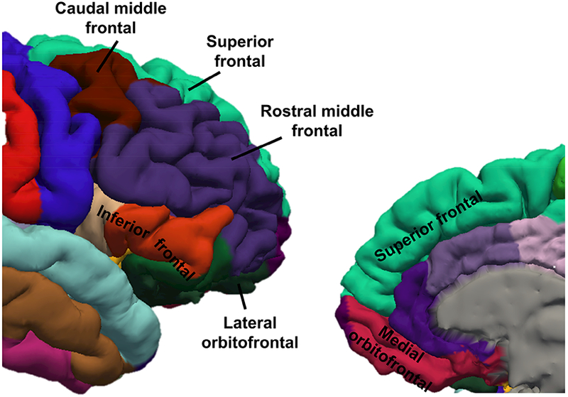
Figure 1. Prefrontal cortical regions of interest (ROIs).

Six ROIs were identified within the prefrontal cortex in each hemisphere for each participant. ROIs were identified by FreeSurfer using Desikan-Killiany atlas and are depicted on restricted lateral (left) and medial (right) views of an example parcellation from the right hemisphere of a 17-year-old female. ROIs depicted: Superior frontal, Rostral middle frontal, Caudal middle frontal, Inferior frontal (a combination of pars orbitalis, pars opercularis, and pars triangularis), Lateral orbitofrontal, Medial orbitofrontal lobes.
