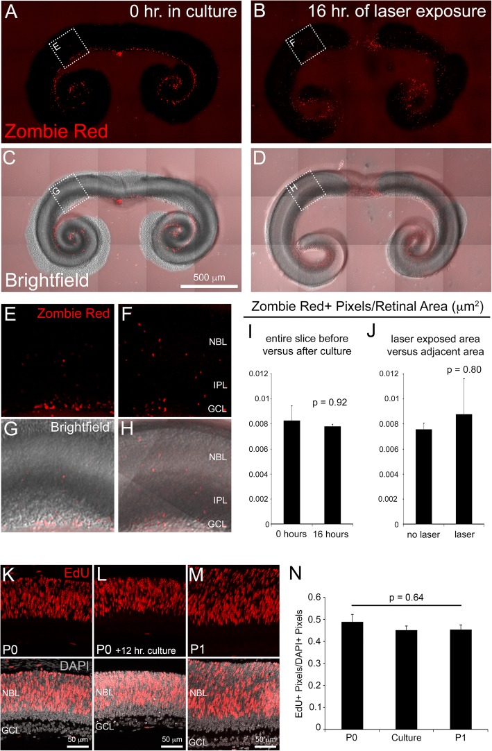
Fig. 2.
Analysis of retinal slice survival and proliferation. Retinal slice cultures stained with Zombie Red™ dye after 0 h (a and c) and 16 h (b and d) in culture. Higher magnifications of boxed region in A-D (E-H). The quantification of Zombie Red+ cells (ZR+ pixels/retinal area) showed no significant differences between 0 h and 16 h in culture (i) or between tissue exposed to laser versus unexposed (j). n = 9 per group. EdU labeling (6-h pulse) and quantification of RPCs in P0 and P1 retinae compared to P0 retinal slices time lapse cultures (k-n). n = 3 per group. Error bars represent SE. Abbreviations: NBL (neuroblastic layer), IPL (inner plexiform layer), GCL (ganglion cell layer)

