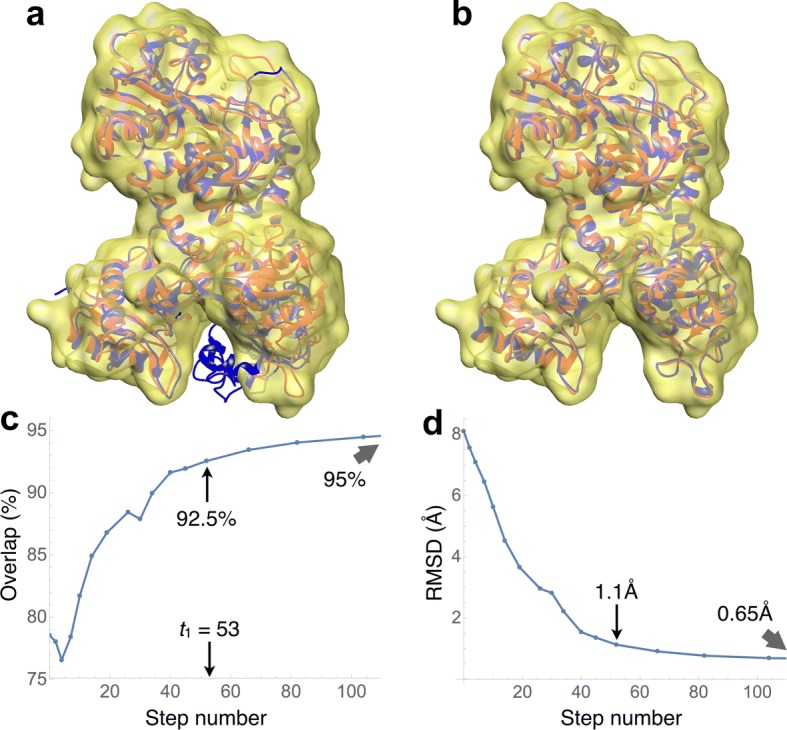
Fig. 4.

Fully flexible fitting of the closed (iron-bound) conformation of lactoferrin (PDB code 1lfg) into a simulated density map (yellow surface) at 7Å resolution. Shown in red is the open conformation (apolactoferrin, PDB code 1lfh), which was used to generate the map. a The closed conformation is shown in blue. This was the starting conformation used for the fitting. b The final conformation of the trajectory is displayed in blue. c Evolution of the overlap values along the trajectory. The indicated value of t1=53 is the warning time, where overfitting is likely to begin. At that time, the overlap was 92.5% d Evolution of the backbone RMSD values along the trajectory. The RMSD was 1.1Å at the t1 time. The stopping time for this case was t2=107
