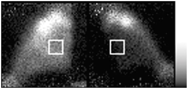
Fig. 2.

Typical posterior-view image of 99mTc PYP in the hind limbs. The left limb was shocked with 1.85 A pulses and 99mTc PYP was injected at 30 min post-shock. The left limb was imaged for 12 min at 105 min post-shock; the right limb was imaged for 12 min at 120 min post-shock. The muscle of the shocked limb (left) shows substantially more uptake than does the muscle of the unshocked limb (right). The white boxes illustrate the typical placement of ROIs over the muscle.
