
Figure 2: Frequencies and numbers of ICAM-1+ neutrophils in lungs from WT and CIRP−/− mice during sepsis.
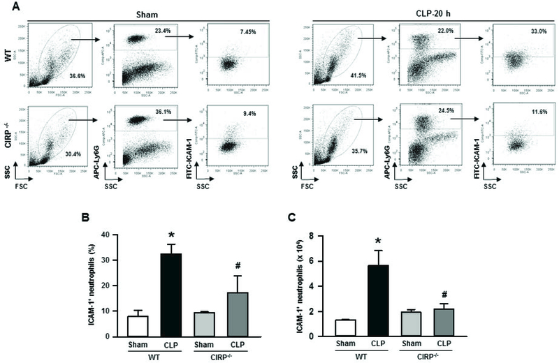
At 20 h of sham or CLP operation, lungs were harvested from WT and CIRP−/− mice. ICAM-1 expression in neutrophils from lung cell lysates was detected by flow cytometry by reacting the cells with APC-Ly6G and FITC-ICAM-1 (CD54) Abs. (A) Representative dot blots of the frequencies of ICAM-1+ neutrophils in lungs from WT and CIRP−/− mice generated from three independent experiments are shown. Diagrammatic presentation of the quantitative mean values of the (B) frequencies and (C) numbers of ICAM-1+ neutrophils in lungs from WT and CIRP−/− mice are shown. Data are expressed as means ± SE (n = 3–5 mice/group) and compared by one-way ANOVA and SNK method (*p < 0.05 vs. WT sham; #p < 0.05 vs. WT CLP). CLP, cecal ligation and puncture; CIRP, cold-inducible RNA-binding protein; ICAM-1, intercellular adhesion molecule.