
Figure 3: Assessment of ICAM-1 expression in BMDN treated with recombinant mouse CIRP in vitro.
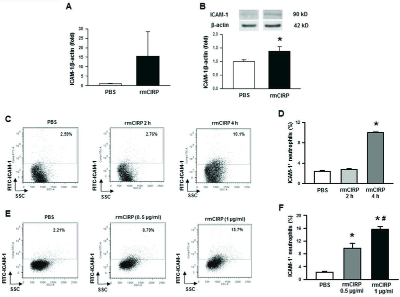
Bone marrows were extracted from tibia and femoral bones of WT mice and BMDN were purified using magnetic beads. (A) A total of 2 × 10⁶ BMDN were treated with rmCIRP (1 µg/ml) for 2 h and then assessed the mRNA levels of ICAM-1 by using real-time PCR. Data are expressed as means ± SE (n = 10 mice/group) and compared by Student’s t test. (B) A total of 2 × 10⁶ BMDN were treated with rmCIRP (1 µg/ml) for 4 h and then assessed the protein levels of ICAM-1 by using Western blot. Data are expressed as means ± SE (n = 7 mice/group) and compared by Student’s t test (*p < 0.05 vs. sham mice). For the detection of surface levels of ICAM-1 expression, a total of 1 × 10⁶ BMDN were stimulated with rmCIRP for various times and doses, followed by the assessment of BMDN by flow cytometry after staining them with APC-Ly6G and FITC-ICAM-1 Abs. (C) Dot blots and their (D) corresponding bar diagram representing the frequencies of ICAM-1+ cells within Ly6G+ populations following treatment of the BMDN with 0.5 µg/ml of CIRP at 2 h and 4 h time points. Data are expressed as means ± SE (n = 9/group) and compared by one-way ANOVA and SNK method (*p < 0.05 vs. PBS). (E) Dot blots and their (F) corresponding bar diagram representing the frequencies of ICAM-1+ cells within Ly6G+ populations following treatment of the BMDN with 0.5 and 1 µg/ml of CIRP for 4 h. Data are expressed as means ± SE (n = 3/group) and compared by one-way ANOVA and SNK method (*p < 0.05 vs. PBS; #p < 0.05 vs. CIRP at 0.5 µg/ml dose). CIRP, cold-inducible RNA-binding protein; ICAM-1, intercellular adhesion molecule; rmCIRP, recombinant murine CIRP; BMDN, bone marrow-derived neutrophils.