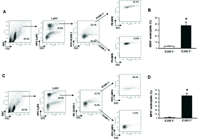
Figure 6: Assessment of iNOS and NETs in ICAM-1+ neutrophils in lungs after sepsis.

At 20 h of sham or CLP operation, lungs were harvested from mice. iNOS and NETs were detected in the single cell suspensions of lung tissues using flow cytometry by reacting the cells with APC-Ly6G, FITC-ICAM-1 (CD54) and PE-iNOS Abs. (A) Representative dot blots of the frequencies of iNOS expressing ICAM-1+ neutrophils in lungs are shown. (B) Diagrammatic presentation of the quantitative mean values of the frequencies of iNOS expressing ICAM-1+ neutrophils in lungs is shown. For the estimation of NETs, a total of 1 × 10⁶ cells isolated from the lung tissues of sham or 20 h after CLP operation were stained with APC-Ly6G, PE-ICAM-1 (CD54) and FITC-MPO Abs and subjected to flow cytometry. (C) Representative dot blots of the frequencies of extracellular MPO expressing ICAM-1+ neutrophils in lungs are shown. (D) Diagrammatic presentation of the quantitative mean values of the frequencies of extracellular MPO expressing ICAM-1+ neutrophils in lungs is shown. Data are expressed as means ± SE (n = 5 mice/group) and compared by Student’s t test (*p < 0.05 vs. ICAM-1−). CLP, cecal ligation and puncture; ICAM-1, intercellular adhesion molecule-1; MPO, myeloperoxidase; iNOS, inducible nitric oxide synthase.
