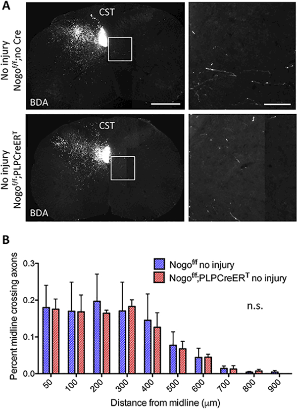
Figure 5. Inducible deletion of Nogo from oligodendrocytes does not affect axon counts in the absence of injury.

(A) BDA-labeled CST axons in cervical sections from Nogof/f;no Cre and Nogof/f;PLPCreERTmice without injury. White boxes depict enlarged regions shown in the right panels. Scale bars = 500 μm (low mag, left), 100 μm (high mag, right). (B) Quantification of the number of BDA-labeled axons at specific distances from the midline contralateral to the main labeled CST normalized to the total labeled CST axon count at the medulla in Nogof/f;no Cre (n=3) and Nogof/f;PLPCreERT(n=3) mice. n.s, not significant by two-way RM ANOVA. Error bar, SEM.
