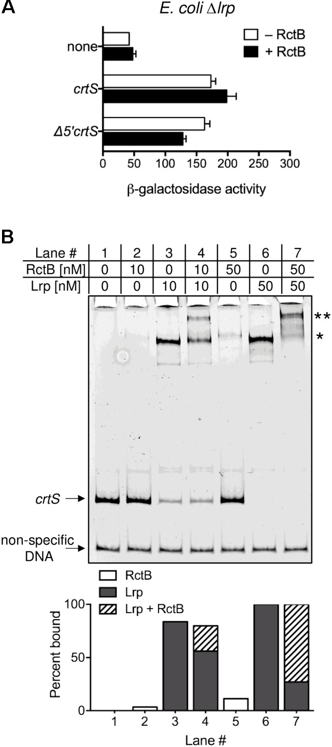
FIGURE 5.

Lrp enhances RctB binding to crtS. (A) Promoter activity of crtS (from PcrtS) was measured as in Figure 2 in E. coli Δlrp (CVC3259) carrying either the empty vector (none, pMLB1109), or the same vector carrying crtS (pBJH235) or its 5′ truncated derivative Δ5′crtS (pPC067). Cells also had either a source of RctB (pRR24) or the corresponding empty vector (pPC020). Note that RctB, which was partially effective in repressing PcrtS in WT, fails to repress the promoter in Δlrp. Data represent mean ± SEM from three biological replicates. (B) EMSA of 5′-FAM labeled crtS DNA (upper arrow) and non-specific DNA (lower arrow) with Lrp and RctB. Both Lrp (lanes three and six) and RctB (lanes two and five) were individually seen to bind crtS specifically. Note that the major Lrp bound band (∗) is super-shifted in the presence of RctB (∗∗). The intensity of the super-shifted band is much higher than the RctB bound band, indicating that RctB binds better to Lrp bound crtS. Shown below are percentages of probe bound to RctB alone (white columns), Lrp alone (gray columns) and, both Lrp and RctB (hatched columns).
