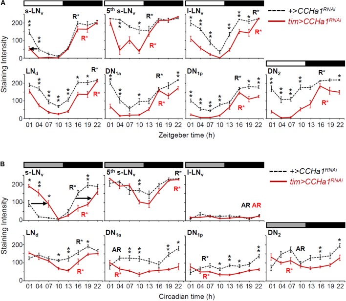
FIGURE 7.
Cyclic expression of the PDP1 clock protein in the CCHa1 knockdown flies. The PDP1 levels (mean ± SEM) in clock neurons were measured at 3-h intervals during LD (A) and DD (B). Circadian time in DD does not indicate the exact subjective time but simply indicates the original zeitgeber time. Hemispheres of 9 different brains were analyzed for each time point. Dashed and colored solid lines indicate the data of the control (UAS-dicer2;+/UAS-CCHa1RNAi) and the RNAi (UAS-dicer2;tim-Gal4/UAS-CCHa1RNAi) strains, respectively. The rhythmicity of PDP1 expression is indicated by R∗ (rhythmic) or AR (arrhythmic), analyzed by CircWave (p < 0.01). (A) The decrease in PDP1 levels in the morning was slightly phase-advanced in the s-LNv neurons (arrow). (B) The PDP1 cycling in the RNAi flies was significantly phase-delayed in the s-LNv neurons compared with that of the control flies (arrows). We performed Mann–Whitney U test followed by Bonferroni correction to examine the effect of CCHa1 knockdown for each time point (∗p < 0.05, ∗∗p < 0.01).

