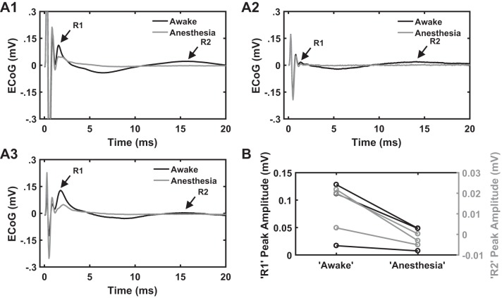
Fig. 10.
A1–A3: cortical evoked potentials (cEP) under anesthesia and awake conditions in response to subthalamic nucleus (STN) deep brain stimulation (DBS) (n = 3). Stimulation settings for electrocorticographic (ECoG) recordings: charge-balanced, symmetric biphasic pulses with a duration of 90 µs/phase, amplitude range of 50–70 µA, and frequency of 50 Hz. Each panel shows cEP response during awake and anesthesia conditions from 1 of the 3 rats. The R2 response is completely suppressed following administration of anesthesia (isoflurane or sevoflurane), whereas the magnitude of R1 response decreased compared with that during the awake condition. B: quantification of R1 and R2 peak amplitudes under anesthesia and awake conditions. Note a decrease in R1 peak amplitude and a complete suppression of R2 response during anesthesia compared with the awake condition across all 3 rats.

