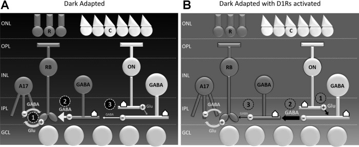
Fig. 7.
Sources of inhibition to rod bipolar cells under dark-adapted conditions and upon D1 receptor activation. A: in dim light, rod bipolar cells receive reciprocal inhibition from A17 amacrine cells (1) and lateral inhibition from wide-field GABAergic amacrine cells (2), as well as narrow-field glycinergic amacrine cells (not shown). When light stimuli of sufficient size are used, the strength of lateral inhibition to rod bipolar cells (2) is diminished by serial inhibition (3). B: when D1 receptors are activated, this circuit is modified at several sites: ON bipolar cell voltage-gated sodium (NaV) channels are inactivated (1), potentially affecting glutamate release onto downstream targets. The strength of serial inhibition is increased (2), either through greater release of GABA by wide-field amacrine cells onto other amacrine cells or the potentiation of GABAA currents in cells that are targets of serial inhibition. GABA release by amacrine cells that mediate lateral inhibition (3) is decreased independent of serial inhibitory influences, possibly through D1-dependent modulation of NaVs or voltage-gated calcium (CaVs). ONL, outer nuclear layer; OPL, outer plexiform layer; INL, inner nuclear layer; IPL, inner plexiform layer; GCL, ganglion cell layer; R, rod photoreceptor; C, cone photoreceptor; RB, rod bipolar cell; Glu, glutamate; ⌂, D1 receptor.

