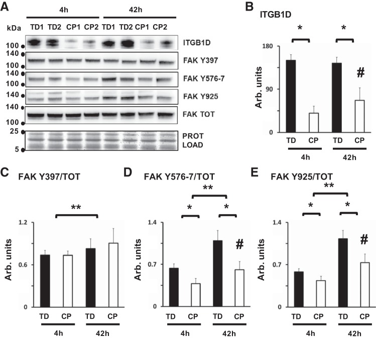
Fig. 5.
Impaired integrin signaling during myoblast fusion and differentiation in cerebral palsy (CP). A: representative Western blots showing differential expression of integrin β-1D (ITGB1D, bands at ~116 kDa), focal adhesion kinase (FAK) phosphorylated at residue Y397 (FAK Y397, ~125 kDa), FAK phosphorylated at Y576/7 residues (FAK Y576/7, ~125 kDa), FAK phosphorylated at Y925 residue (FAK Y925, ~125 kDa), total FAK protein levels (FAK TOT, ~125 kDa), and total protein (bands in the 5–25 kDa range). Protein analysis was performed in CP and typically developing (TD) cell preparations, 4 h (4h) and 42 h (42h) after high- to low-serum medium switch. B: protein quantification of ITGB1D; *CP vs. TD, P < 0.0001; #CP after 42 h, P = 0.01, analysis by two-way ANOVA (n = 8/group). C: protein quantification for FAK Y397, normalized over FAK TOT; **4 h vs. 42 h, P = 0.025, analysis by two-way ANOVA (n = 6/group). D: protein quantification for FAK Y576/7, normalized over FAK TOT; *CP vs. TD, P < 0.0001, **4 h vs. 42 h, P < 0.0001, #CP after 42 h, P = 0.034, analysis by two-way ANOVA (n = 6/group). E: protein quantification for FAK Y925, normalized over FAK TOT, *CP vs. TD, P < 0.0001, **4 h vs. 42 h, P < 0.0001, #CP after 42 h, P = 0.009, analysis by two-way ANOVA (n = 6/group). Only significant P values are reported.

