Fig. 2.
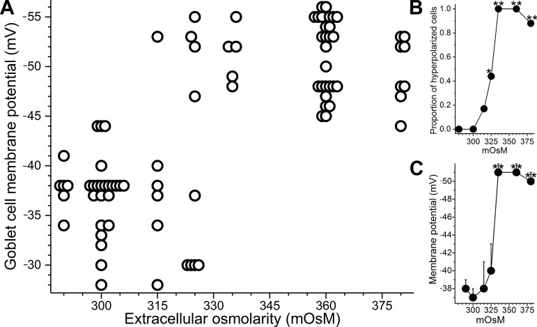
Effect of extracellular osmolarity on the membrane potential of conjunctival goblet cells. A: resting membrane potentials of goblet cells located in freshly isolated rat conjunctival specimens exposed for 10 to 30 min to solutions of various osmolarities. To aid visualization, symbols are displaced slightly when multiple goblet cells had identical membrane potentials. B: proportion of sampled goblet cells with resting membrane potentials more negative than −44 mV. C: mean membrane potentials at the 7 osmolarities assayed. *P = 0.0078; **P < 0.0001.

