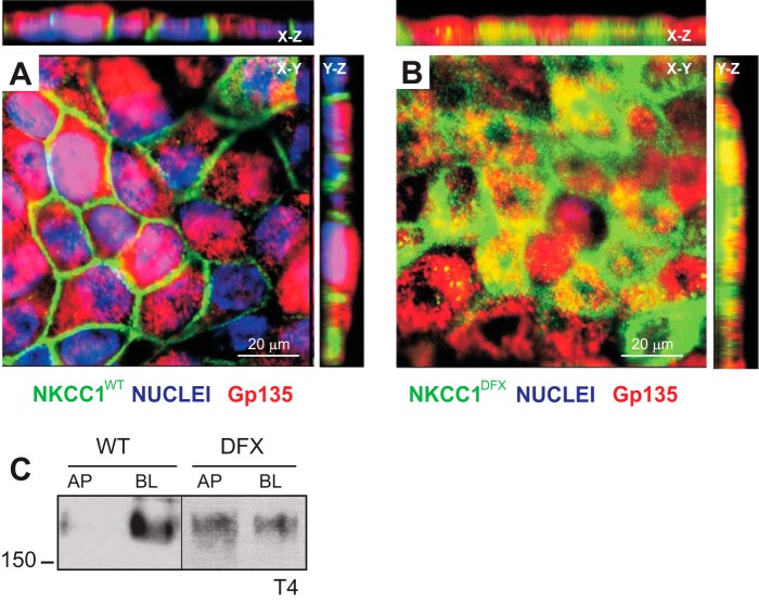
Fig. 5.
NKCC1-DFX mistrafficks from basolateral membrane to subapical pole and apical membrane. A and B: MDCK cells stably transfected with eGFP-NKCC1-WT (A) and eGFP-NKCC1-DFX (B) were polarized on Transwell filters for 5 days. Cells were then fixed and stained with anti-GFP and Gp135/podocalyxin. Z-sections were captured at 5-μm intervals on the Zeiss LSM 880 microscope. Bars, 20 μm. C: polarized MDCK cells expressing NKCC1-WT and NKCC1-DFX were selectively biotinylated on the apical membrane or basolateral membrane. Cell lysates were prepared, and equal amounts of protein were immunoprecipitated with streptavidin agarose beads and analyzed by Western blotting. AP, apical; BL, basolateral; eGFP, enhanced green fluorescent protein; MDCK, Madin-Darby canine kidney; NKCC1, Na+-K+-2Cl− cotransporter-1; WT, wild type.

