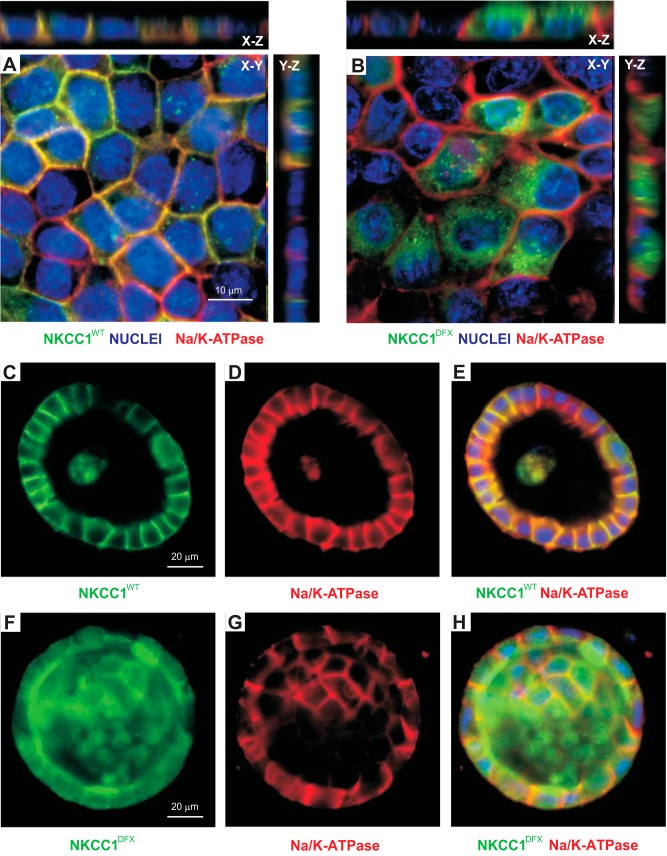
Fig. 11.
NKCC1-DFX does not affect the polarity of MDCK cells. A and B: MDCK cells stably transfected with NKCC1-WT (A) or NKCC1-DFX (B) were grown on coverslips for 5 days and immunostained with antibodies to GFP (GFP-NKCC1) and the α-subunit of the Na-K-ATPase. C–H: localization of Na-K-ATPase (red) and NKCC1 (green) in MDCK cells transfected with eGFP-NKCC1-WT (C–E) or eGFP-NKCC1-DFX (F–H), and grown in 3-D extracellular matrix. Scale bars, 10 μm or 20 μm. eGFP, enhanced green fluorescent protein; MDCK, Madin-Darby canine kidney; NKCC1, Na+-K+-2Cl− cotransporter-1; WT, wild type.

