Fig. 15.
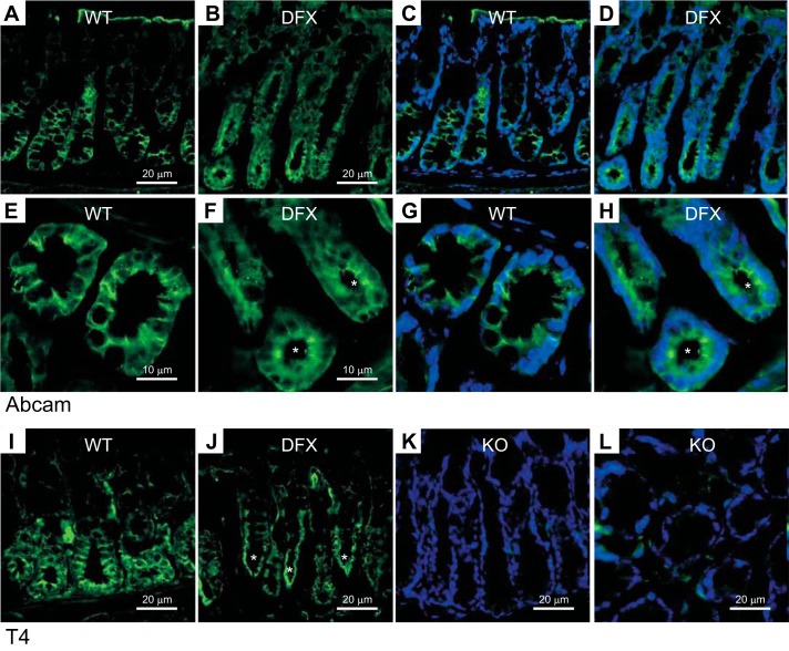
NKCC1 is mistrafficked to the apical/subapical membrane in NKCC1WT/DFX mouse colon. A and B: representative fluorescence micrographs showing NKCC1 (green) expression in colon sections from NKCC1WT/WT (A) and NKCC1WT/DFX (B) mice using the Abcam NH2-specific NKCC1 antibody. Scale bars, 20 μm. C and D: same panels as A and B with DAPI staining nuclei. E and F: high magnification of NKCC1WT/WT and NKCC1WT/DFX colon crypt regions. G and H: same panels as E and F with DAPI staining nuclei. I: T4 (carboxyl-terminus-specific) antibody was used to stain NKCC1WT/WT colon sections. J: T4 antibody was used to stain NKCC1WT/DFX colon sections. K and L: T4 antibody was used to stain NKCC1DFX/DFX (NKCC1-KO) colon sections. Scale bars, 20 μm. KO, knockout; NKCC1, Na+-K+-2Cl− cotransporter-1; WT, wild type. Asterisks indicate NKCC1 signal at apical pole.

