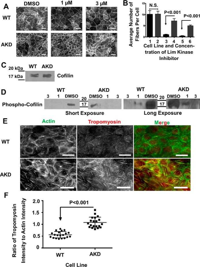
Figure 6.

Stress fibers in α-actinin–depleted cells are less susceptible to cofilin-mediated disassembly and contain more tropomyosin. A, micrographs of the basal surface of WT and AKD cells treated with DMSO and 1 or 3 μm Lim kinase inhibitor. Scale bars are 20 μm. B, graph of the average number of filaments per cell, ±95% confidence interval, in WT and AKD cells as a function of the amount of Lim kinase inhibitor. Bar 1 is WT control cells; bar 2 is AKD control-treated cells; bar 3 is WT cells treated with 1 μm inhibitor; bar 4 is AKD cells treated with 1 μm inhibitor; bar 5 is WT cells treated with 3 μm inhibitor; and bar 6 is AKD cells treated with 3 μm inhibitor. C, developed Western blotting probing cofilin levels in WT and AKD cells demonstrating no significant change in protein levels. D, developed Western blots of cells treated with DMSO or Lim kinase inhibitor and probed for phospho-cofilin. Both a short and long exposure are given. and 20 μg of protein was added to each lane. E, micrographs of WT or actinin–depleted cells stained for actin and tropomyosin. Scale bars are 20 μm. F, graph of the ratio of tropomyosin to actin along stress fibers in WT and depleted cells. The mean ratio was significantly greater in the depleted cells.
