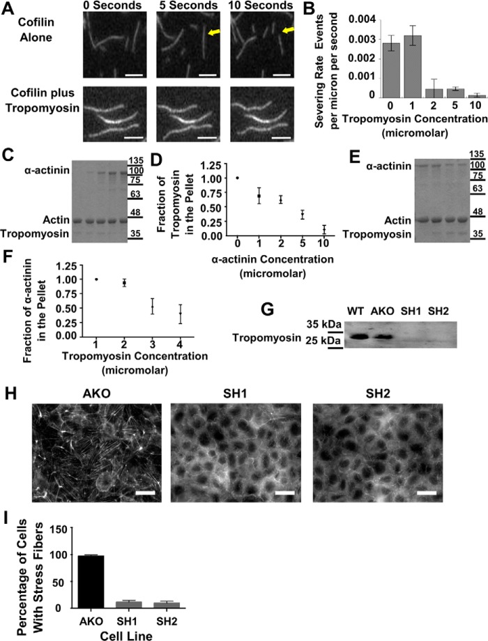
Figure 7.
Tropomyosin provides stability to actin fibers both in vitro and in vivo, and α-actinin competes for access to actin filaments in vitro. A, consecutive micrographs from an in vitro severing assay of actin filaments either in the presence or absence of tropomyosin. The arrows point to a filament that severs in the presence of cofilin. Scale bars are 5 μm. B, graph of the severing rate of actin filaments via cofilin as a function of tropomyosin concentration. C, Coomassie Blue-stained gel of the pellets from spin-down assays where tropomyosin concentration was maintained and α-actinin concentration was increased from left to right. D, graph of the amount of tropomyosin in the pellet as a function of actinin concentration from repeated experiments. E, Coomassie Blue-stained gel of the pellets from spin-down assays where actinin concentration was maintained and tropomyosin concentration was increased from left to right. F, graph of the amount of actinin in the pellet as a function of tropomyosin concentration from repeated experiments. G, Western blotting probing tropomyosin in WT, actinin knockout, and actinin knockout transfected with tropomyosin ShRNA1 or ShRNA2. H, micrographs of actinin knockout cell line or actinin knockout transfected with SH1 or SH2 and stained for actin. I, graph quantifying the percentage of cells with stress fibers as a function of transfection with SH1 or SH2.

