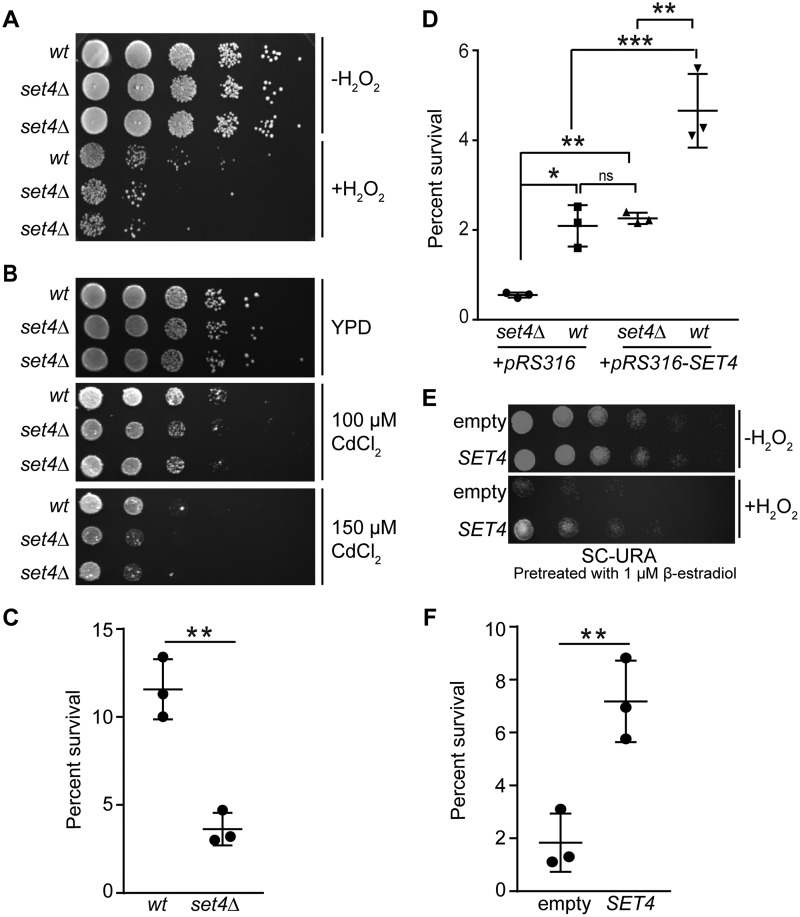
Figure 2.
Set4 promotes survival during oxidative stress. A, 10-fold serial dilutions of WT (yEG001) and set4Δ (yEG322 and yEG325) strains grown to mid-log phase in YPD or YPD with 4 mm H2O2 for 30 min. B, 10-fold serial dilutions of WT (yEG001) and set4Δ (yEG322 and yEG325) strains spotted on YPD, YPD with 100 μm CdCl2, or YPD with with 150 μm CdCl2. C, percentage survival calculated from cfu of WT (yEG001) and set4Δ (yEG325) strains following treatment with 4 mm H2O2 for 30 min. The scatter plot shows the mean and S.D. (error bars) from three biological replicates. An unpaired t test was used to determine statistical significance. D, percentage survival of WT and set4Δ cells (yEG638, yEG639, yEG640, and yEG641) carrying either the empty pRS316 vector or pRS316-SET4, with SET4 under the control of its endogenous promoter, calculated from cfu on SC-URA following treatment with 20 mm H2O2 for 30 min. One-way analysis of variance was performed, followed by Turkey's post hoc test to determine statistical significance. E, 10-fold serial dilutions of strains carrying either pMN3 (empty) or pMN3-SET4 (yEG372 and yEG375, respectively) that were pretreated with 1 μm β-estradiol for 4–5 h, followed by exposure to 20 mm H2O2 for 30 min. F, percentage survival calculated from cfu of cells grown as in E, except that 50 nm β-estradiol was used to induce SET4, and plated on SC-URA. An unpaired t test was used to determine statistical significance. For all panels, asterisks represent p values as follows: *, p ≤ 0.05; **, p ≤ 0.01; ***, p ≤ 0.001. ns, not significant.

