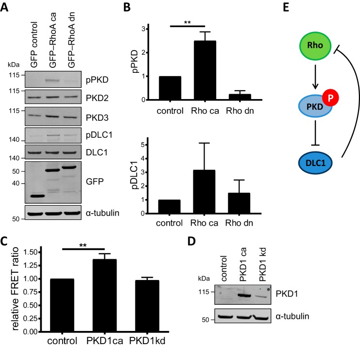
Figure 1.
Reciprocal activation of PKD and RhoA. A, Flp-In GFP-DLC1 cells were transiently transfected with vectors encoding GFP, ca GFP-RhoA, or dn GFP-RhoA. Six hours post-transfection, GFP-DLC1 expression was induced with doxycycline. The next day, cells were lysed, and lysates were analyzed by immunoblotting. B, band intensities from three independent experiments were quantified and normalized to the loading control and control sample. Shown are the means with error bars depicting ±S.E. C, HEK293T cells were transiently cotransfected with vectors encoding ca or kinase-dead (kd) PKD1 and a RhoA FRET biosensor (27). The next day, cells were lysed, and the FRET ratio was analyzed. Shown are the mean FRET ratios from three independent experiments normalized to the control. Shown are the means with error bars depicting ±S.E. D, cell lysates from C were analyzed by immunoblotting. E, positive feedback hypothesis in which Rho activates PKD, which phosphorylates and inactivates the RhoGAP DLC1, to support further Rho activation. Data in B and C were analyzed by one-way analysis of variance followed by Dunnett's multiple comparisons test. Only statistically significant changes are indicated. **, p < 0.01.

