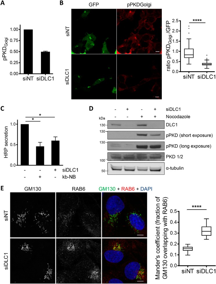
Figure 6.
DLC1 depletion impairs protein secretion. A and B, 2 days post-siRNA transfection, HEK293T cells were transfected with the vector encoding the PKD reporter. A, the next day, cells were lysed, and lysates were analyzed by immunoblotting. The pPKDGolgi reporter signal was normalized to the GFP signal. Shown are the mean values of two independent experiments. The error bar indicates ±S.E. B, the next day, cells were fixed and stained with an antibody reactive with the phosphorylated reporter. PKD activity at the Golgi was determined by ratiometric imaging of 92 cells from two independent experiments. Scale bar, 10 μm. ****, p < 0.0001 (two-sample t test). C, Flp-In T-REx 293 FLAG-HRP cells were transfected with the indicated siRNAs. Expression of FLAG-HRP was induced with doxycycline. The next day, the medium was replaced with serum-free medium containing either kb-NB or DMSO. The supernatant was collected after 5 h for HRP measurements. Shown are the means with error bars depicting ±S.E. *, p < 0.05 (one-sample t test). D, U2OS cells were transfected with the indicated siRNAs. After 3 days, the cells were treated with nocodazole or DMSO for 10 min and lysed. The lysates were analyzed by immunoblotting with the indicated antibodies. E, U2OS cells were transfected with the indicated siRNAs. After 3 days, the cells were fixed and stained with antibodies specific for GM130 and RAB6. The images shown are maximum intensity projections of several confocal sections. Colocalization of GM130 and RAB6 (n = 11; N = 3) was analyzed with ImageJ. Scale bar, 10 μm. ****, p < 0.0001 (two-sample t test). DAPI, 4′,6-diamidino-2-phenylindole.

