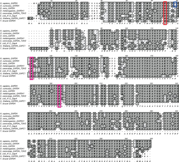
Figure 1.
GAPDH protein alignment. The three strictly conserved histidine residues are highlighted in red (human, His-53) and magenta (human, His-111 and His-179). In blue is His-57 (human) found in human and rabbit GAPDH. His-111 and His-179 are found in stable secondary structure motifs, whereas His-53 and His-57 are located on a flexible loop at the interprotomer interface.

