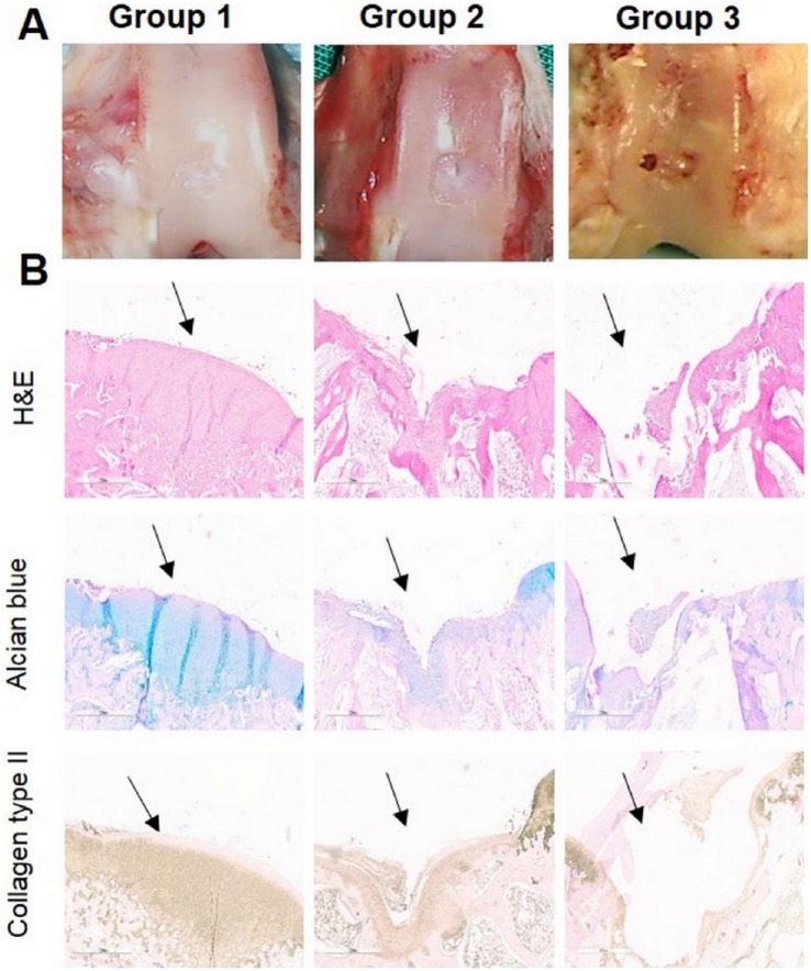
Figure 3.
Representative images of macroscopic (A), histological and immunohistochemical observation (B) of chondral defects at 12 weeks postoperatively repaired by microfracture + PRP (Group 1), transplantation of autologous ADSCs + PRP (Group 2), and without any treatment (Group 3). Arrows show the defect. Scale bar = 600 µm (magnification 20×).

